Chapter 4 — Fourier in hydraulics
The cochlea, slowly
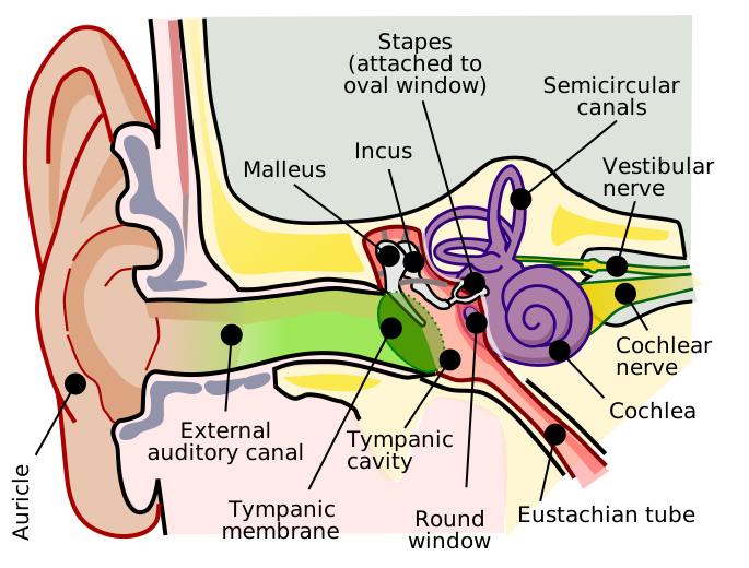
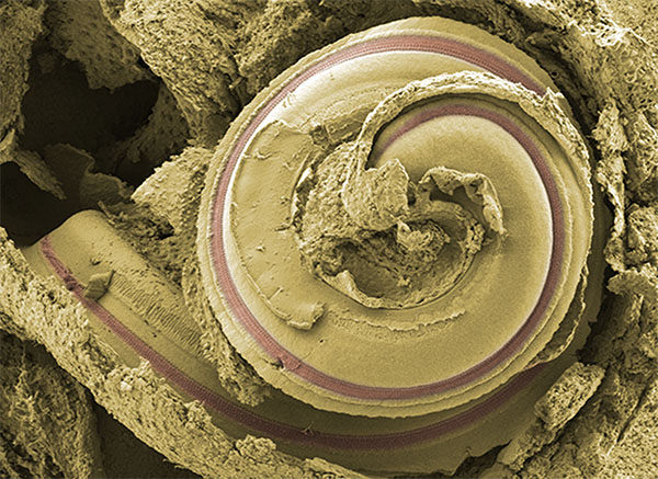
By the time “Hey Dr. Miles!” reaches the oval window, it has already become a mechanical motion — the stapes acting as a small piston driven by the ossicular lever we built in movement 4. From here on, the signal lives in fluid. The cochlea is where that fluid motion becomes a frequency map; nowhere else in the auditory system does so much computational work get done without a single neuron firing.
This chapter takes the journey at a deliberate pace. There are seven sub-sections: the geometry first (4.1), then the stiffness gradient of the basilar membrane (4.2), the wave that travels along it (4.3), the place map (4.4), the active feedback that sharpens it (4.5), the inner hair cells that read it out (4.6), and finally what leaves through the auditory nerve (4.7). It is a lot. It is also where the essay either earns the math reader’s attention or doesn’t, and it is not rushed.
The sub-sections are listed in the top nav and walk through the cochlea in order. The math accumulates: 4.1 is pure anatomy; 4.2 introduces the local damped oscillator and its closed-form response; 4.3 introduces the fluid-coupled wave equation; 4.4 introduces the Greenwood place map; 4.5 introduces the active amplifier; 4.6 introduces the Boltzmann transduction curve; 4.7 ties it together. Each equation comes with a collapsible derivation from first principles.
For the underlying anatomical reference, the 3-D ear model linked below is the canonical visual — open it in a tab and consult it whenever a structure I introduce here is unclear. The inline 2-D figures in this essay show only what we need for the current physics argument; the 3-D model is where to go for the full structural picture.